Instead, electrons scanned across the sample are absorbed, and secondary electrons are emitted from the sample. Scanning electron microscopy is a little bit like reflected light microscopy, but in this case it is not electrons from the original beam that return to the detector to make up an image. The end results is that electrons are able to pass through the clear and lightly stained regions of the sample, whereas stained parts of the sample scatter the electrons and appear dark on the image. This is done with a solution of heavy metal salts such as Uranium or Lead. Because the sample is so thin, it needs to be stained to improve contrast. On a light microscope we might use a 20 micrometer thick sample with some success, on a TEM the sample is usually about 60 nanometres thick (a micrometer is 1 millionth of a meter, a nanometer is one billionth of a meter). TEM is very similar to transmitted light microscopy, only in this case it is a beam of electrons, rather than a beam of light, which is passed through the sample. These are called Transmission Electron Microscopy (TEM) and Scanning Electron Microscopy (SEM). Just like with light microscopy, where we have transmitted and reflected light microscopy, we also have two types of electron microscopy. By comparison, an electron microscope can be used to see much smaller structures within biological and material specimens. Electron microscopyĪs we have already seen, the light microscope can be used to look at single cells and we can even see something of their internal structure. If we need to look at very small structures and we don't mind killing the cell in the process, we can learn much more about fine structure with the electron microscope. This is a bit like the difference between trying to pick up something while wearing a thick pair of gloves, and then trying the same thing with a pair of tweezers. On the down side, we are limited in how much we can magnify a sample because the wavelength of light is quite long, as compared to the wavelength of the electron beam used in electron microscopy. Light microscopy is a very useful tool in biology because the light used to image the sample does not tend to kill it. By adding several dyes, which stain different parts of the cell, we can increase the contrast in the sample and see more of the biological structures we're interested in by seeing less of the structures surrounding them. the dye DAPI only stains a cell's nucleus). This is done by putting a dye onto the sample, which becomes attached to a specific part of the cell (e.g. Fluorescence microscopy is a light microscopy technique that dramatically improves contrast. Although we could magnify the image to make it look bigger, we wouldn't actually be able to see anything much at all because there is no contrast. Sometimes, the sample is so thin or translucent that not enough light is absorbed to see a difference (imagine covering your window with pieces of selotape - it would be difficult to see the seloptape on a sunny day). By using a magnifying lens, we can use this approach to see structures in fairly small samples, such as cells.
#TRANSMISSION LIGHT MICROSCOPY WINDOWS#
Because the sample is made up of different structures, light travelling through it is absorbed in different ways (in much the same way that when your windows are covered in gunk you see darker and lighter patches depending on how much sunlight is absorbed and how much gets through).

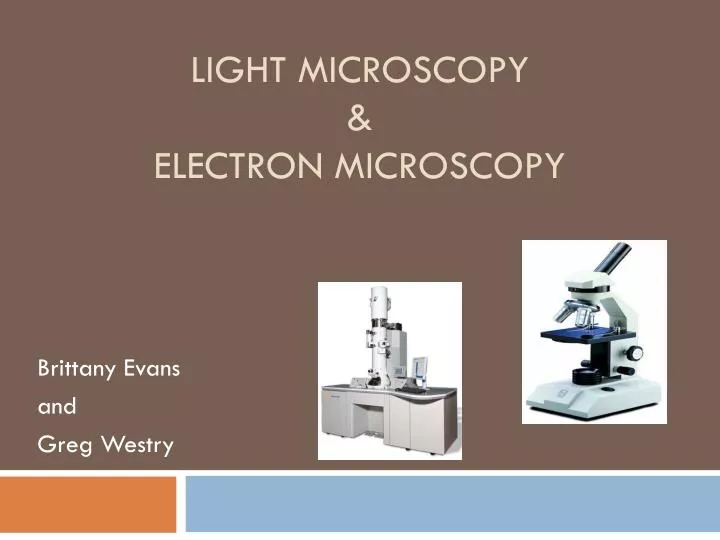
This can be shone through the sample (this is called transmitted light microscopy) or bounced off the surface (reflected light microscopy). The most common type of light microscopy involves shining a regular tungsten filament bulb, a bit like you have in your light fittings at home, onto the sample. Reference the following links for helpful introductions to microscopy: Light microscopy Both these technologies are available in the facility. There are 2 main types of microscope - LIGHT microscopes and ELECTRON microscopes. The Cell Imaging Core is primarily equipped to benefit research scientists within the Faculty of Medicine & Dentistry at the University of Alberta, and is also available to users from other academic institutes and commercial companies.


 0 kommentar(er)
